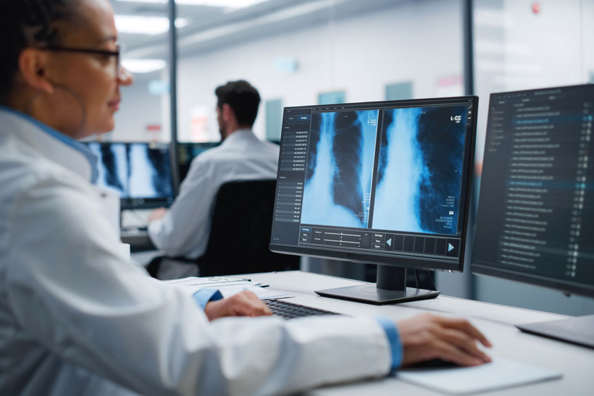
Medical imaging saves millions of lives each year. It helps doctors to detect and diagnose a wide range of diseases, from cancer and appendicitis to stroke and heart disease.
Artificial intelligence (AI) is supporting real-world solutions across the healthcare space, including medical imaging. With help from AI, imaging technicians don’t even need to rely on their eyes alone to identify potential concerns.
Let’s take a look at some of the biggest medical imaging technology trends shaping the industry.
What are the new advancements in medical imaging?
Artificial and augmented intelligence
The market for artificial intelligence in medical imaging is slated to reach $14.2 billion by 2032 (up from $762 million in 2022). With hundreds of AI technologies in development—including machine learning (ML), natural language processing, and augmented intelligence—vendors will need to prove customer ROI in a competitive setting. They’ll also need to overcome challenges related to data security and transparency to earn authorization from the Food and Drug Administration.
It’s worth the extra effort, though.
AI has the potential to revolutionize the advanced medical imaging industry. Already, AI is being used to help physicians sift through large volumes of scans and return diagnostic insights, giving them more time to oversee treatments and work directly with patients. AI-driven analytics and predictive analytics can even improve the accuracy and speed of decision-making around treatment options.
Take these integrations, for example:
- Google’s DeepMind can read 3D retinal OCT scans and diagnose 50 different ophthalmic conditions with 99% accuracy. It can detect indicators of eye disease. It can also rank patients by urgency and recommend treatment. These capabilities could cut down on the delay between scan and treatment. This allows patients to get sight-saving treatments in time.
- iCAD’s “ProFound AI” is a solution for digital breast tomosynthesis (DBT). It helps radiologists to view each tissue layer and thereby detect cancer up to 8 percent sooner on average. This can reduce radiologists’ time spent reading breast scans by more than 50%.
- Siemens Healthineers and Intel partnered to explore how AI can improve cardiac MRI diagnostics. Currently, cardiologists need to segment many different parts of the heart in their imaging—an incredibly time-consuming task. This AI-enabled instant segmentation technology enables specialists to see more patients each day.
While true artificial intelligence emulates human-like thinking without intervention, augmented intelligence systems in healthcare aim to enhance human processes by improving monotonous or burdensome physician workflows.
In medical imaging, augmented intelligence improves collaboration between radiologists and oncologists by reducing repetitive data-entry tasks and automatically appending electronic health records (EHRs) with imaging and diagnostic data. Augmented intelligence can also be used to improve image quality, recommend customized imaging protocols based on patient history, and generate automated reports.
Virtual and augmented reality & 3D medical imaging
Virtual reality (VR) and augmented reality (AR) technologies are still finding their footing. The VR/AR market continues to underperform, even as Apple just announced its entry into the space and Meta continues to dump billions each quarter into the tech. But the real transformative value of VR is in healthcare, where it’s being used to treat patients and help providers more effectively deliver care.
Chief among these new applications is 3D medical imaging. As powerful as MRIs and CT scans are now, their 2D renders demand physicians to imagine a spatial dimension that they can’t actually see. New augmented reality technologies, like EchoPixel True 3D, make it possible for physicians to create a 3D image of MRIs. They can then examine the image with a typical VR headset, like Apple’s consumer-focused Vision Pro.
Physicians can rotate, zoom in, or make cross sections of the 3D image using peripheral pointing devices and other tools. This enables better visualization and planning before a procedure. Some VR platforms even allow physicians to practice surgeries using these images, or create physical models using 3D printers.
AR is like VR in that it creates three-dimensional images, but projects them into the real world using spatial computing and a special headset. Companies like Proprio are using machine learning and AR to help surgeons see through obstacles and blockages that may impede a high-risk operation.
Nuclear imaging
In nuclear imaging, a patient ingests radioactive materials called radiotracers (or radiopharmaceuticals) before a medical imaging scan. During a scan, a camera focuses on where the radioactive material concentrates. These types of scans are particularly helpful when diagnosing internal conditions like thyroid disease, cancer, and Alzheimer’s disease.
For example, amyloid PET imaging can help predict Alzheimer’s progression. This scan determines whether patients with memory issues have excessive amyloid plaques, which are an indicator of Alzheimer’s disease. Before amyloid PET imaging, these plaques could only be detected by examining the brain during autopsies. Earlier detection means more effective treatment and better patient outcomes.
The EXPLORER Total-body PET/CT Scanner began moving into hospitals for the first time in 2018 with a hefty price tag of $10 million. It uses a much lower dose of radiotracer (18F-FDG) than traditional scanners while also producing higher quality images in less time than earlier models.
Wearables
Wearable medical devices aren’t just monitoring heart rates and insulin levels anymore (although those functions remain critical). A few notable devices show considerable promise for radiology and diagnostic imaging.
In May 2023, scientists and engineers at the Washington University of Medicine in St. Louis received a National Institutes of Health grant to commercialize their wearable brain-imaging device. Instead of using loud, heavy fMRI technology, the device—known as a diffuse optical tomography, or HD-DOT, instrument—uses LEDs to project infrared light into the head and detectors to measure any light passing through. HD-DOT makes it possible to observe patient brain activity during routine activities, or to monitor patients experiencing unusual neurological events like seizures.
Later that year, MIT researchers announced they had developed wearable ultrasound scanners with applications in bladder and kidney disease, breast cancer detection, and more. The flexible devices can be tuned to take images of nearly any organ, suggesting use cases in home health care, diagnostic imaging, and preventive care management.
Learn more
The medical imaging market is constantly evolving. If you’re operating in or selling into it, the right data can help you understand where it’s headed, how your competitors are reacting, and what you can do to get ahead.
When you’re ready to get the full view of the medical imaging market, sign up for a free trial and see how our healthcare commercial intelligence helps you find new opportunities for commercial success.


